Llame a
(414) 643-9000
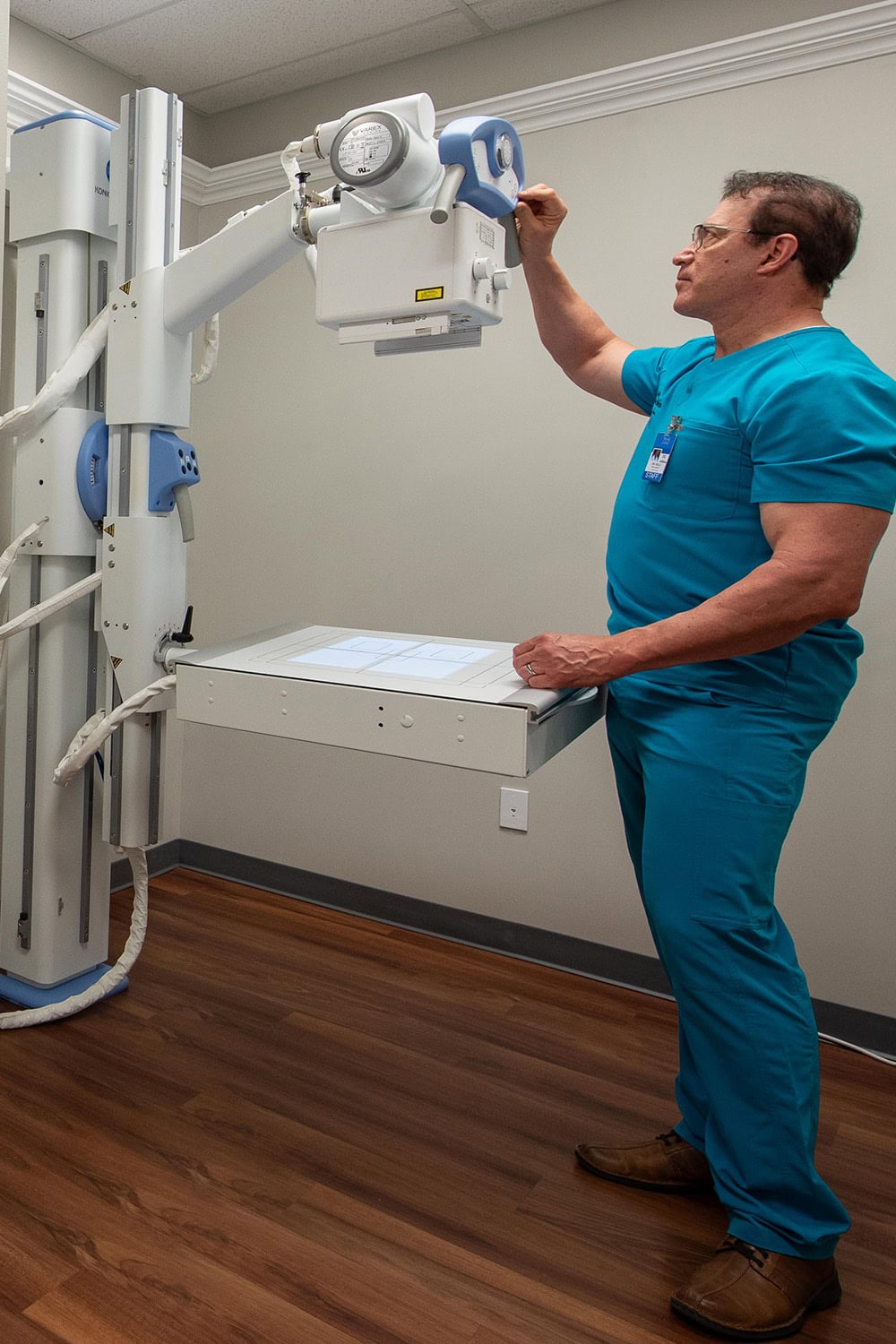
Spine and Joint Institute of Milwaukee cuenta con uno de los únicos sistemas de cinerradiografía de todo Wisconsin, que ahora presta servicio a la región sudeste de Wisconsin. Este innovador sistema permite a nuestro departamento radiológico captar vistas completas tanto de los huesos largos como de toda la columna vertebral, lo que permite una evaluación precisa de fracturas, microfracturas y otras lesiones. A diferencia de las radiografías tradicionales, nuestro sistema proporciona una visión holística de la columna vertebral en condiciones naturales de carga de peso, así como de movimiento, si es necesario, lo que ayuda a la detección precoz de la rotura de ligamentos y lesiones de tejidos blandos.
A esto se añade el movimiento de la parte del cuerpo lesionada, que mostrará mucho más que una radiografía capturada fija. Con la radiografía de movimiento, se puede mostrar la detección de alteraciones en los ligamentos que pueden pasar desapercibidas fácilmente en el estudio de resonancia magnética. Con nuestro sistema, podemos detectar las anormalidades a través del estudio de movimiento y encontrar las alteraciones, documentarlo, e informar objetivamente, dando al representante legal, compañía de seguros o médico tratante, las razones detrás del dolor persistente de alguien que sigue sin ser diagnosticado.
Nuestro sistema de cinerradiografía digital ofrece resultados de imágenes inmediatos que se muestran en un sofisticado panel de pantalla plana a través de una interfaz informatizada. Esta tecnología reduce el tiempo de inactividad, facilita el análisis rápido por parte de nuestros radiólogos médicos y permite incluir fácilmente las imágenes en informes completos. Comprometido con la curación de nuestros pacientes, nuestro instituto garantiza que todas las necesidades radiológicas se satisfagan con precisión y eficacia.
Si se somete a un examen de Radiografía Digital Dinámica, puede esperar lo siguiente:
No olvide comentar los resultados de su radiografía digital con su profesional sanitario, ya que puede ofrecerle más información y recomendaciones basadas en los resultados.
La tecnología de radiografía digital funciona de forma similar a la radiografía tradicional, pero existen algunas diferencias clave en la forma en que se capturan y procesan las imágenes.
Al igual que las radiografías convencionales, las radiografías digitales utilizan radiación electromagnética para penetrar en el cuerpo. Esta radiación interactúa de forma diferente con los distintos tejidos, creando sombras que forman la imagen.
En lugar de película, las radiografías digitales utilizan un detector digital para capturar la imagen. Existen dos tipos principales de detectores digitales: los detectores de panel plano (FPD) y los detectores de radiografía computarizada (CR). Los FPD convierten directamente los rayos X en señales eléctricas, mientras que los detectores CR utilizan una placa de fósforo de almacenamiento que capta la energía de los rayos X y se explora para producir una imagen digital.
Cuando los rayos X atraviesan el cuerpo, los distintos tejidos los absorben a ritmos diferentes. El detector digital capta estas diferencias en la absorción de la radiación y las convierte en una señal electrónica.
La señal electrónica del detector se envía a un ordenador, donde se procesa y convierte en una imagen digital. Esta imagen puede manipularse para una visualización óptima ajustando el contraste, el brillo y otros parámetros.
Una vez procesada, la imagen radiográfica digital puede visualizarse en el monitor de un ordenador o en otro dispositivo de visualización. Esto permite a los radiólogos y otros profesionales sanitarios interpretarla inmediatamente.
Las radiografías digitales pueden almacenarse electrónicamente, lo que elimina la necesidad de almacenar películas físicas. También pueden transmitirse fácilmente por vía electrónica, lo que permite la consulta a distancia y el intercambio entre profesionales sanitarios.
La tecnología de rayos X digital ofrece varias ventajas con respecto a las radiografías de película tradicionales, como una adquisición de imágenes más rápida, una menor exposición a la radiación y una mayor flexibilidad en la manipulación y el almacenamiento de imágenes.

Quiero darles las gracias. Gracias a ellos, hoy puedo trabajar. Debido a la lesión que tuve, me dolían el hombro, el codo y la mano. Gracias a los médicos y especialistas de Spine and Joint, mi rehabilitación fue rápida. Gracias de todo corazón, muchas bendiciones.

Empecé en el Spine and Joint Institute con mucho dolor en el hombro y no podía ni moverlo. Con la ayuda de la cirugía y mucha terapia todo mejoró notablemente. Ahora puedo mover el brazo gracias a las terapias y al trato amable de todas las personas que trabajan aquí. Realmente recomiendo Spine and Joint Institute.
Hoy junio ll terminé mi tratamiento y quiero agradecer a Spine and Joint porque gracias a ellos vuelvo a mis rutinas normales sin dolor. Gracias a todo el personal que fueron muy atentos y amables en cada terapia que tuve y me ayudaron a sanar rápidamente.
Quiero agradecer al Spine and Joint institute el buen servicio que me dieron ya que con su ayuda mi hombro y muñeca mejoraron mucho.

Gracias al Spine and Joint Institute y al Dr. Kelly mis hombros quedaron muy bien. Después de la cirugía hicieron un gran trabajo conmigo. Gracias también a Priscila y a todo el equipo, estoy muy bien.
Después de un examen, nuestros pacientes son remitidos a nuestro departamento de Rayos X, donde nuestro técnico de Rayos X licenciado y certificado toma las radiografías específicas ordenadas por el médico. Una vez terminadas, el médico revisa los detalles de las radiografías y las evalúa con el paciente. Buscará fracturas, luxaciones o lesiones ligamentosas que puedan aparecer en la radiografía. A continuación, las radiografías se envían a nuestro radiólogo médico certificado, que las lee y elabora un informe detallado de los hallazgos, normalmente en un plazo de 24-48 horas. Una vez que el informe está completo, proporcionamos una copia al paciente y revisamos los resultados con ellos.
En Spine and Joint Institute of Milwaukee, puede estar seguro de que sus necesidades radiológicas serán satisfechas. Con un sistema digital de alta resolución para tomas fijas, o rayos X de movimiento cineradiográfico, su lesión será documentada y diagnosticada adecuadamente para que podamos recuperarle. Entendemos que su capacidad para ganarse la vida se ha visto afectada. Su informe radiológico y las radiografías le indicarán cuándo es el momento adecuado para volver al trabajo, ya sea con restricciones o a jornada completa. Nunca le enviaremos de vuelta antes de lo necesario.
En Spine and Joint Institute of Milwaukee, puede contar con nosotros para obtener las radiografías y el informe radiológico que necesita, para documentar objetivamente su lesión e indicarle cuándo puede volver al trabajo.